WORKSHOPS
Limited Spaces Available
Places at workshops are only available to fully registered delegates. Places are limited, and on a first-in-first-served basis. You can reserve and pay for your workshop during the registration process
WORKSHOPS
Advanced Fluorescence Workshop FULLY SUBSCRIBED
NO SPACES AVAIL.ABLE
The workshop will cover an introduction to FRET, SRRF, spectral unmixing and deconvolution in the morning session. The afternoon will be an introduction to Leica SP5, How to measure a point spread function, How to acquire and process spectral images, Blind deconvolution, Spectral unmixing with reference spectra vs blind unmixing, Super-resolution by SRRF demo by Coherent Scientific. FRET demo.
Limited to 6 persons – participants are encouraged to bring a sample to test – please check with the workshop organiser for excitation wavelengths available and provide details on your test specimen. Sorry we can’t deal with PC2 samples in our lab unless they are fixed and dead.
Transport from Hamilton is included in this workshop cost.

Dr Lloyd Donaldson is a leading microscopist specialising in plant anatomy and ultrastructure,... More

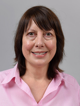
Jacqui Ross is a staff member in the Biomedical Imaging Research Unit at The University of... More

Dr Robert Woolley has a broad scientific research career spanning a host of scientific... More
Introduction to Electron Backscatter Diffraction (EBSD) and Transmission Kikuchi Diffraction (TKD)
EBSD has become a well-established technique for microstructural characterisation in the SEM or FIB. The crystallographic data obtained can be used in the measurement of crystal orientation, misorientation, grain size distribution, texture, strain analysis, recrystallised / deformed fractions and grain boundary characterisation. The technique is used in a broad span of applications in many fields including metals & materials analysis and processing, microelectronics and earth sciences.
More recent developments in SEM-based diffraction have led to the technique of transmission kikuchi diffraction (TKD) which overcomes some of the resolution limitations of conventional EBSD enabling characterisation of materials at the nanoscale.
This one day workshop is an introduction to EBSD & TKD and will cover the following topics:
- The basic physics and how an EBSD system works
- What information can we get from EBSD and what kind of experiments can be performed
- Hardware, calibration and tips for setting up the SEM for EBSD and TKD
- Sample preparation and why it is important
- Data presentation and analysis
- The benefits of combined EDS and EBSD
- Applications of EBSD and TKD

Julie Sheffield-Parker is a Director of Nanospec Pty Ltd, distributors for Oxford Instruments... More
OME (Open Microscopy Environment) - Imaging workflows in OMERO
The Open Microscopy Environment (OME) is an open-source software project that develops tools that enable access, analysis, visualization, sharing and publication of biological image data. OME supports more than 150 image data formats across many imaging modalities including fluorescence microscopy, high-content screening, whole-slide imaging and biomedical imaging.
OME supports and visualizes multi-channel 3D timelapse images. OMERO is an open source, enterprise software platform developed by OME for image data management and analysis. OMERO is used in 1000s of institutions worldwide managing, sharing, analysing and publishing imaging datasets.
This workshop will cover all of the main functions of OMERO. We will demonstrate data import, organisation, viewing, searching, annotation and publishing. After we cover the basics of OMERO, we will demonstrate manual data processing and automated processing workflows using a range of open source applications running alongside OMERO. We will demonstrate how to integrate a variety of processing tools with OMERO such as ImageJ/Fiji, R and CellProfiler and how to programmatically generate output ready for publication.
This workshop is designed for researchers at all levels who work with data from digital microscopes or other imaging systems. The workshop includes presentations and hands-on session. Prior knowledge in microscopy, scripting and data analysis is not required.
Any student/researcher dealing with scientific images is more than welcome to join this workshop.

Petr Walczysko joined the OME project in October 2012 as a software specialist for testing and... More
A romp through the meadow of Image Analysis into the land of Machine Learning
Have a plan of attack, it saves grief later on; consider your options, fancy might be tempting, but simple has its attractions too; and if thresholding has driven you to distraction, then the basics of machine learning might be the image analysis yoga-mat for you.
Image analysis isn’t a process to be done in isolation, irrespective of which method is chosen. Simply having a great method to extract information from an image is pointless if the image has been taken, selected, or processed in the wrong way. Numbers can lie.
This workshop probably won’t give you a customised method to analyse your images on the day, but it should give you the tools to think through an appropriate method to give accurate and reliable results when you return to your own comfortable lab.
The tools of choice will be Fiji (ImageJ) for general analysis techniques and using it in conjunction with Ilastik for the fabulous world of machine learning. Some overview will also be made of 3DSlicer, which is useful for 3D volumes, meshes, 3D alignments and suchlike. All are open source and run on Linux, OS X and Windows.

Andrew works with confocal and light microscopes, and the X-ray µCT. Consequently, he has... More
POST CONFERENCE WORKSHOP
STED (Stimulated Emission Depletion) Workshop
Friday 15th of November 10:00 – 17:00
Location – Biomedical Imaging Research Unit, The University of Auckland
The workshop starts in the morning with an introduction to STED and related technologies like RESCue STED, DyMin and MINFIELD. In the afternoon we will be able to use the Abberior Instruments STEDYCON system to image samples in confocal mode and then achieve 30 nm resolution in STED mode.
Limited to 6 persons – participants are encouraged to bring their own samples. Please check with the workshop organisers for the right dyes/ secondary antibodies and provide details on your test specimen.
There is no charge for attending the workshop but participants must arrange their own transport to the workshop location. However, there will be a number of conference delegates travelling to Auckland on Thursday 14 November, who may be able to offer to share transport.
Contact Jacqui Ross at jacqui.ross@auckland.ac.nz to register for the workshop.

Workshop Facilitators

|
Jacqueline Ross |

|
Dr Carola Thoni |


